The only thing that's worse than losing a bird is not knowing why, and being therefore unable to help others in similar circumstances.
With that in mind, I have translated the preliminary results of Tigga's necropsy (culture results still to come) so that we can use them as a learning resource. I have added a glossary of terms, and some pictures to illustrate them.
TIGGA'S NECROPSY:Background information:Feral squeaker, about 10 weeks old.
Found: Portugal, 18th June 2010.
Weight: 162 g, poor condition.
Droppings: bright green, oily-looking.
Medicated with Flagyl and Zooserine (Tetracycline chlorhydrate 90mg/g, Chloramphenicol 66.5mg/g, and Furadolizone 15mg/g).
Only lived a further one and a half days.
Macroscopic examination:Very thin.
Pronouced anemia in the mucose, subcutaneous tissue and entrails.
Liver colouration: lead green.
Kidneys: Discoloured, showing nodular whitish lesions about 0.1 cm in diameter, resembling a granuloma.(1)
Small intestine containing yellowish paste, showing a greener colour in the large intestine.
Samples taken:Heart, lung, liver, gizzard, kidneys, small intestine, sciatic nerve.
Microscopic examination:Lung: Acute congestion and pronounced hemosiderosis(2). Presence of rounded or oblong, sometimes multinucleated cells with giant oval nuclei, resembling syncytia(3).
Heart: Pronounced congestion and hemosiderosis. Mononucleated inflammatory infiltrate(4) rich in macrophages (5), with very few plasmocytes(6) and lymphocytes (7) -- focal pericarditis(8). A round structure was observed in the thrombus (9) of the lumen (10) of a coronary artery, and another one in the adventitia (11) of the same artery. In the lumen of the right ventricle there was another round structure with a closer similarity to a protozoan resembling Balantidium (12).
Liver: Presence of elongated, oval, basophilic (13) structures resembling megabacteria. Disorganisation of the hepatic sinusoids (14) with hepatocyte (15) vacuolisation (16) and pronounced hemosiderosis. Presence of mixed inflammatory infiltrate rich in lymphocytes, plasmocytes and heterophile antibodies (17) in the hepatic sinusoids -- hepatitis of apparently infectious origin.
Gizzard: Without significant tissue alteration.
Small intestine: There were oval or rounded eosinophilic (18) structures in the lumen of the small intestine, corresponding to an infectious agent of unknown provenance. Scaling of the lining of the intestinal mucosa and total necrosis of the intestinal wall.
Large intestine: Scaling of the lining of the intestinal mucosa and total necrosis of the wall. The same infectious agents present in the small intestine were found here, albeit in smaller numbers.
Kidneys: Coagulative necrosis (19) and scaling of the renal mucosa, apparently aggravated by autolysis (20).
Sciatic nerve: Mixed inflammatory infiltrate located in the perineural (21) area of the sciatic nerve.
Glossary of terms:(1) Granuloma: a roughly spherical mass of immune cells that forms when the immune system attempts to wall off substances that it perceives as foreign but is unable to eliminate. Such substances include infectious organisms such as bacteria and fungi, as well as other materials such as keratin and suture fragments.
Infectious diseases characterised by the presence of granulomas include tuberculosis, leprosy, histoplasmosis, cryptococcosis, cat-scratch disease, sarcoidosis, Crohn's disease, pneumocystis pneumonia and aspiration pneumonia.
Granuloma:

(2) Hemosiderosis: a form of iron overload disorder resulting in the accumulation of hemosiderin (an iron deposit found within cells, as opposed to circulating in blood, and which causes organ damage).
Hemosiderin deposition in the lungs is often seen after diffuse alveolar hemorrhage. Mitral stenosis can also lead to pulmonary hemosiderosis. Hemosiderin deposition in the liver is a common feature of hemochromatosis and is the cause of liver failure in the disease. Hemosiderin deposition in the brain is seen after bleeds from any source.
Hemosiderosis (brown staining):

(3) Syncytium: a large cell-like structure filled with cytoplasm containing many nuclei. A syncytium can form in two ways: by incomplete cell division or by the fusion of thousands of individual cells.
A syncytium is also the normal cell structure for many fungi and algae. More primitive fungi may contain multiple nuclei in a true syncytium.
Syncytium (formation of):

(4) Infiltrate: Material or solution deposited in the interstices of a tissue or substance.
(5) Macrophage: A type of white blood cell that ingests foreign material. Macrophages are key players in the immune response to foreign invaders such as infectious microorganisms.
Blood monocytes migrate into the tissues of the body and there differentiate (evolve) into macrophages.
Macrophages help destroy bacteria, protozoa, and tumor cells. They also release substances that stimulate other cells of the immune system.
Macrophage (illustration):

(6) Plasmocytes: White blood cells that produce large volumes of antibodies.
Plasmocyte:

(7) Lymphocytes: Small white blood cells (leukocytes) that are responsible for immune responses. There are two main types of lymphocytes: B cells and T cells. The B cells make antibodies that attack bacteria and toxins while the T cells attack body cells themselves when they have been taken over by viruses or have become cancerous.
Lymphocytes secrete products (lymphokines) that modulate the functional activities of many other types of cells and are often present at sites of chronic inflammation.
Lymphocyte:

(8) Pericarditis is an inflammation of the sac that surrounds the heart. It can be caused by an infection (viral, bacterial or fungal), a heart attack, trauma, a tumour, cancer, a connective tissue disease, kidney failure or adverse reaction to some medications.
(9) Thrombus: blood clot.
Thrombus:

(10) Lumen: interior space of a blood vessel or other tubular structure.
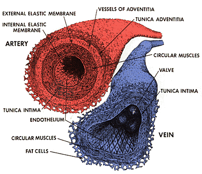
(11) Adventitia: the outermost layer of connective tissue covering an organ or blood vessel.
(Tunica) Adventitia:

(12)
Balantidium coli: a protozoan which causes an infection called Balantidiasis. Symptoms can be local due to involvement of the intestinal mucosa, or systemic in nature and include either diarrhea or constipation.
Treatment: Balantidiasis can be treated with carbarsone, tetracycline, or diiodohydroxyquin.
Balantidium coli:

(13) Basophilic describes the appearance of structures seen in histological sections which take up basic dyes. A Basophil granulocyte stains dark blue upon H&E staining. The most common such dye is haematoxylin. The structures usually stained are those that contain nucleic acid such as the cell nucleus and ribosomes.
Basophile (purple):

(14) Hepatic sinusoids: the intralobular vascular supply system, lined by endothelial cells and stellate macrophages.
Hepatic sinusoids:

(15) Hepatocytes: cells which make up most of the liver's cytoplasmic mass. These cells are involved in: protein synthesis; protein storage; the transformation of carbohydrates; the synthesis of cholesterol, bile salts and phospholipids; detoxification, modification, and excretion of exogenous and endogenous substances.
The hepatocyte also initiates formation and secretion of bile.
Hepatocytes:

(16) Vacuolisation: the state of having become filled with vacuoles.
Vacuolised hepatocytes:

(17) Heterophile antibodies: those which react with antigens not responsible for their production.
(18) Eosinophilic: describes the appearance of cells and structures seen in histological sections that take up the staining dye eosin. This is a bright-pink dye that stains the cytoplasm of cells.
Eosinophilic erythrocytes (pink):

(19) Coagulative necrosis: a type of necrosis in which the affected cells or tissue are converted into a dry, dull, fairly homogeneous eosinophilic mass without nuclear staining, as a result of the coagulation of protein as occurs in an infarct. Microscopically, the necrotic process involves chiefly the cells, and remnants of histologic elements (e.g., elastin, collagen, muscle fibres) may be recognizable, as well as ghosts of cells and portions of cell membranes; may be caused by heat, ischemia (a restriction in blood supply, or local anemia resulting from vasoconstriction), and other agents that destroy tissue, including enzymes that would continue to alter the devitalised cellular substance.
Coagulative necrosis (discoloured, centre):

(20) Autolysis: more commonly known as self-digestion, refers to the destruction of a cell through the action of its own enzymes.
(21) Perineural: around a nerve or group of nerves.


